Next: Mechanoreceptors Up: 11.2 The Physiology of Previous: Middle ear Contents Index
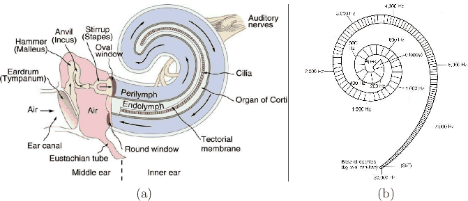
The inner ear contains both the vestibular organs, which were covered in Section 8.2, and the cochlea, which is the sense organ for hearing. The cochlea converts sound energy into neural impulses via mechanoreceptors. This is accomplished in a beautiful way that performs a spectral decomposition in the process so that the neural impulses encode amplitudes and phases of frequency components.
 |
Figure 11.5 illustrates its operation. As seen in Figure 11.5(a), eardrum vibration is converted into oscillations of the oval window at the base of the cochlea. A tube that contains a liquid called perilymph runs from the oval window to the round window at the other end. The basilar membrane is a structure that runs through the center of the cochlea, which roughly doubles the length of the tube containing perilymph. The first part of the tube is called the scala vestibuli, and the second part is called the scala tympani. As the oval window vibrates, waves travel down the tube, which causes the basilar membrane to displace. The membrane is thin and stiff near the base (near the oval and round windows) and gradually becomes soft and floppy at the furthest away point, called the apex; see Figure 11.5(b). This causes each point on the membrane to vibrate only over a particular, narrow range of frequencies.
Steven M LaValle 2020-11-11