
Next: Impact on perception Up: 8.2 The Vestibular System Previous: Sensing linear acceleration Contents Index
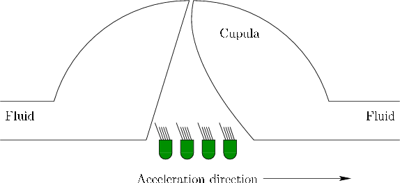 |
The semicircular canals use the same principle as the otolith organs. They measure acceleration by bending cilia at the end of hair cells. A viscous fluid moves inside of each canal. A flexible structure called the cupula blocks one small section of the canal and contains the hair cells; see Figure 8.7. Compare the rotation of a canal to the merry-go-round. If we were to place a liquid-filled tube around the periphery of the merry-go-round, then the fluid would remain fairly stable at a constant angular velocity. However, if angular acceleration is applied, then due to friction between the fluid and the tube (and also internal fluid viscosity), the fluid would start to travel inside the tube. In the semicircular canal, the moving fluid applies pressure to the cupula, causing it to deform and bend the cilia on hair cells inside of it. Note that a constant angular velocity does not, in principle, cause pressure on the cupula; thus, the semicircular canals measure angular acceleration as opposed to velocity. Each canal is polarized in the sense that it responds mainly to rotations about an axis perpendicular to the plane that contains the entire canal.