Next: Sensing linear acceleration Up: 8.2 The Vestibular System Previous: 8.2 The Vestibular System Contents Index
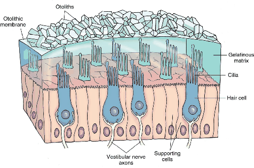
Figure 8.4 shows the location of the vestibular organs inside of the human head. As in the cases of eyes and ears, there are two symmetric organs, corresponding to the right and left sides. Figure 8.3 shows the physiology of each vestibular organ. The cochlea handles hearing, which is covered in Section 11.2, and the remaining parts belong to the vestibular system. The utricle and saccule measure linear acceleration; together they form the otolith system. When the head is not tilted, the sensing surface of the utricle mostly lies in the horizontal plane (or ![]() plane in our common coordinate systems), whereas the corresponding surface of the saccule lies in a vertical plane that is aligned in the forward direction (this is called the sagittal plane, or
plane in our common coordinate systems), whereas the corresponding surface of the saccule lies in a vertical plane that is aligned in the forward direction (this is called the sagittal plane, or ![]() plane). As will be explained shortly, the utricle senses acceleration components
plane). As will be explained shortly, the utricle senses acceleration components ![]() and
and ![]() , and the saccule senses
, and the saccule senses ![]() and
and ![]() (
(![]() is redundantly sensed).
is redundantly sensed).
The semicircular canals measure angular acceleration. Each canal has a diameter of about ![]() to
to ![]() mm, and is bent along a circular arc with a diameter of about
mm, and is bent along a circular arc with a diameter of about ![]() to
to ![]() mm. Amazingly, the three canals are roughly perpendicular so that they independently measure three components of angular velocity. The particular canal names are anterior canal, posterior canal, and lateral canal. They are not closely aligned with our usual 3D coordinate coordinate axes. Note from Figure 8.4 that each set of canals is rotated by
mm. Amazingly, the three canals are roughly perpendicular so that they independently measure three components of angular velocity. The particular canal names are anterior canal, posterior canal, and lateral canal. They are not closely aligned with our usual 3D coordinate coordinate axes. Note from Figure 8.4 that each set of canals is rotated by ![]() degrees with respect to the vertical axis. Thus, the anterior canal of the left ear aligns with the posterior canal of the right ear. Likewise, the posterior canal of the left ear aligns with the anterior canal of the right ear. Although not visible in the figure, the lateral canal is also tilted about
degrees with respect to the vertical axis. Thus, the anterior canal of the left ear aligns with the posterior canal of the right ear. Likewise, the posterior canal of the left ear aligns with the anterior canal of the right ear. Although not visible in the figure, the lateral canal is also tilted about ![]() away from level. Nevertheless, all three components of angular acceleration are sensed because the canals are roughly perpendicular.
away from level. Nevertheless, all three components of angular acceleration are sensed because the canals are roughly perpendicular.
 |
Steven M LaValle 2020-11-11