
Next: Medical motivation Up: 13.4 Brain-Machine Interfaces Previous: 13.4 Brain-Machine Interfaces Contents Index
 |
The goal of devices that measure neural activity is to decipher the voluntary intentions and decisions of the user. They are usually divided into two categories: non-invasive (attaching sensors to the skin is allowed) and invasive (drilling into the skull is allowed).
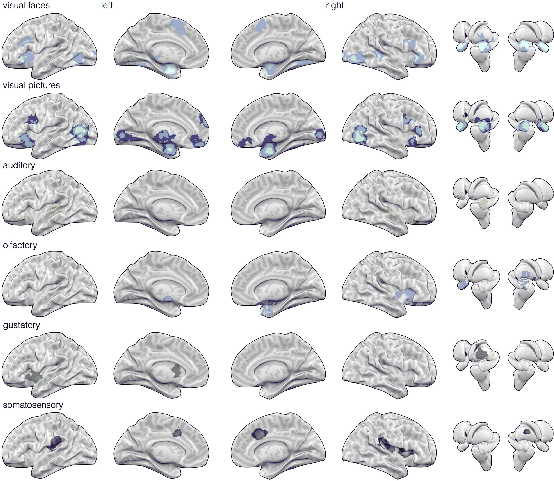
First consider the non-invasive case, which is by far the most appropriate for humans. The most accurate way to measure full brain activity to date is by functional magnetic resonance imaging (fMRI), which is shown in Figure 13.10. This is related to MRI, which most people are familiar with as a common medical scanning method. Ordinary MRI differs in that it provides an image of the static structures to identify abnormalities, whereas an fMRI provides images that show activities of parts of the brain over time. Unfortunately, fMRI is too slow, expensive, and cumbersome for everyday use as a VR interface [175]. Furthermore, users must remain rigidly fixed, and sometimes they ingest a dye that increases contrast due to variations in blood flow.
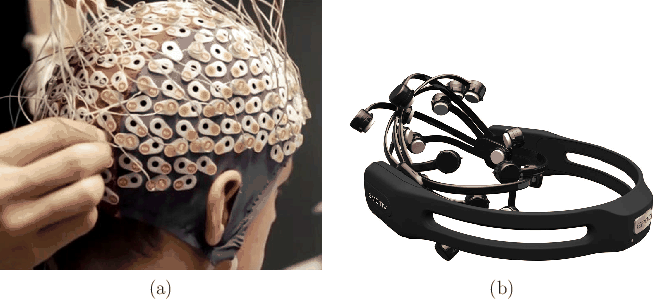 |
Thus, the most common way to measure brain activity for BMI is via electroencephalogram (EEG), which involves placing electrodes along the scalp to measure electrical field fluctuations that emanate from neural activity; see Figure 13.11. The signal-to-noise ratio is unfortunately poor because the brain tissue, bone, and skin effectively perform low-pass filtering that destroys most of the signal. There is also significant attenuation and interference with other neural structures. The transfer rate of information via EEG is between 5 and 25 bits per second [175,359]. This is roughly equivalent to one to a few characters per second, which is two orders of magnitude slower than the average typing rate. Extracting the information from EEG signals involves difficult signal processing [282]; open-source libraries exist, such as OpenVibe from INRIA Rennes.
For the invasive case, electrodes are implanted intracranially (inside of the skull). This provides much more information for scientists, but is limited to studies on animals (and some humans suffering from neural disorders such as Parkinson's disease). Thus, invasive methods are not suitable for the vast majority of people as a VR interface. The simplest case is to perform a single-unit recording for a particular neuron; however, this often increases the number of required trials because the neural response typically switches between different neurons across trials. As the number of neurons increases, the problem of deciphering the thoughts becomes more reliable. Numerous recordings could be from a single site that performs a known function, or could come from multiple sites to help understand the distributed processing performed by the brain [175].