
Next: Photoreceptor density Up: 5.1 From the Cornea Previous: Parts of the eye Contents Index
 |
 |
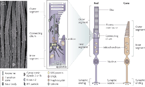
The retina contains two kinds of photoreceptors for vision: 1) rods, which are triggered by very low levels of light, and 2) cones, which require more light and are designed to distinguish between colors. See Figure 5.2. To understand the scale, the width of the smallest cones is around ![]() nm. This is quite close to the wavelength of visible light, implying that photoreceptors need not be much smaller. Each human retina contains about
nm. This is quite close to the wavelength of visible light, implying that photoreceptors need not be much smaller. Each human retina contains about ![]() million rods and
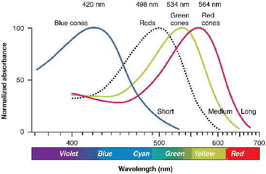
million rods and ![]() million cones that are densely packed along the retina. Figure 5.3 shows the detection capabilities of each photoreceptor type. Rod sensitivity peaks at
million cones that are densely packed along the retina. Figure 5.3 shows the detection capabilities of each photoreceptor type. Rod sensitivity peaks at ![]() nm, between blue and green in the spectrum. Three categories of cones exist, based on whether they are designed to sense blue, green, or red light.
nm, between blue and green in the spectrum. Three categories of cones exist, based on whether they are designed to sense blue, green, or red light.
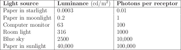 |
Photoreceptors respond to light levels over a large dynamic range.
Figure 5.4 shows several familiar examples. The
luminance is measured in SI units of candelas per square meter, which
corresponds directly to the amount of light power per area. The range
spans seven orders of magnitude, from ![]() photon hitting a
photoreceptor every
photon hitting a
photoreceptor every ![]() seconds up to
seconds up to ![]() photons per receptor
per second. At low light levels, only rods are triggered. Our
inability to distinguish colors at night is caused by the inability of
rods to distinguish colors. Our eyes may take up to 35 minutes to
fully adapt to low light, resulting in a monochromatic mode called
scotopic vision. By contrast, our cones become active in
brighter light. Adaptation to this trichromatic mode, called
photopic vision, may take up to ten minutes (you have
undoubtedly noticed the adjustment period when someone unexpectedly
turns on lights while you are lying in bed at night).
photons per receptor
per second. At low light levels, only rods are triggered. Our
inability to distinguish colors at night is caused by the inability of
rods to distinguish colors. Our eyes may take up to 35 minutes to
fully adapt to low light, resulting in a monochromatic mode called
scotopic vision. By contrast, our cones become active in
brighter light. Adaptation to this trichromatic mode, called
photopic vision, may take up to ten minutes (you have
undoubtedly noticed the adjustment period when someone unexpectedly
turns on lights while you are lying in bed at night).
 |
Steven M LaValle 2020-11-11